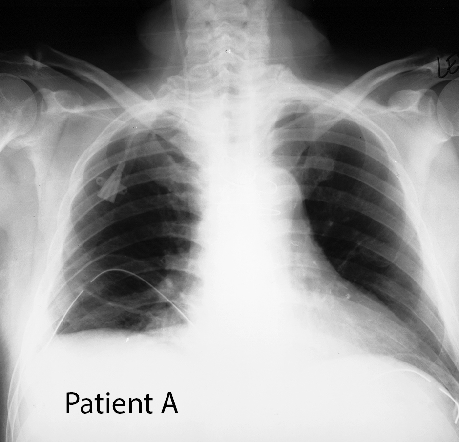
These cases can be reviewed individually or with other students before or after mini-consolidation session on Chest Imaging.
For a brief review of types of radiological imaging, with narrated slides, click this LINK.
Learning Objectives:
After review of online resources and in class mini-consolidation, students will be able to:
1. List the common sites for placement of a central line (CVL), and identify the expected course and ideal location for the catheter tip on chest radiographs (frontal and lateral), using knowledge of venous anatomy of the chest and of how patient anatomy is displayed on radiography.
2. List two causes of absence of lung markings on a chest radiograph, and describe the space in which abnormal air would be located in each clinical setting, as well as potential effects on change in position of the heart on venous return.
Thorax Case 1 Thorax Case 2