

Case 3
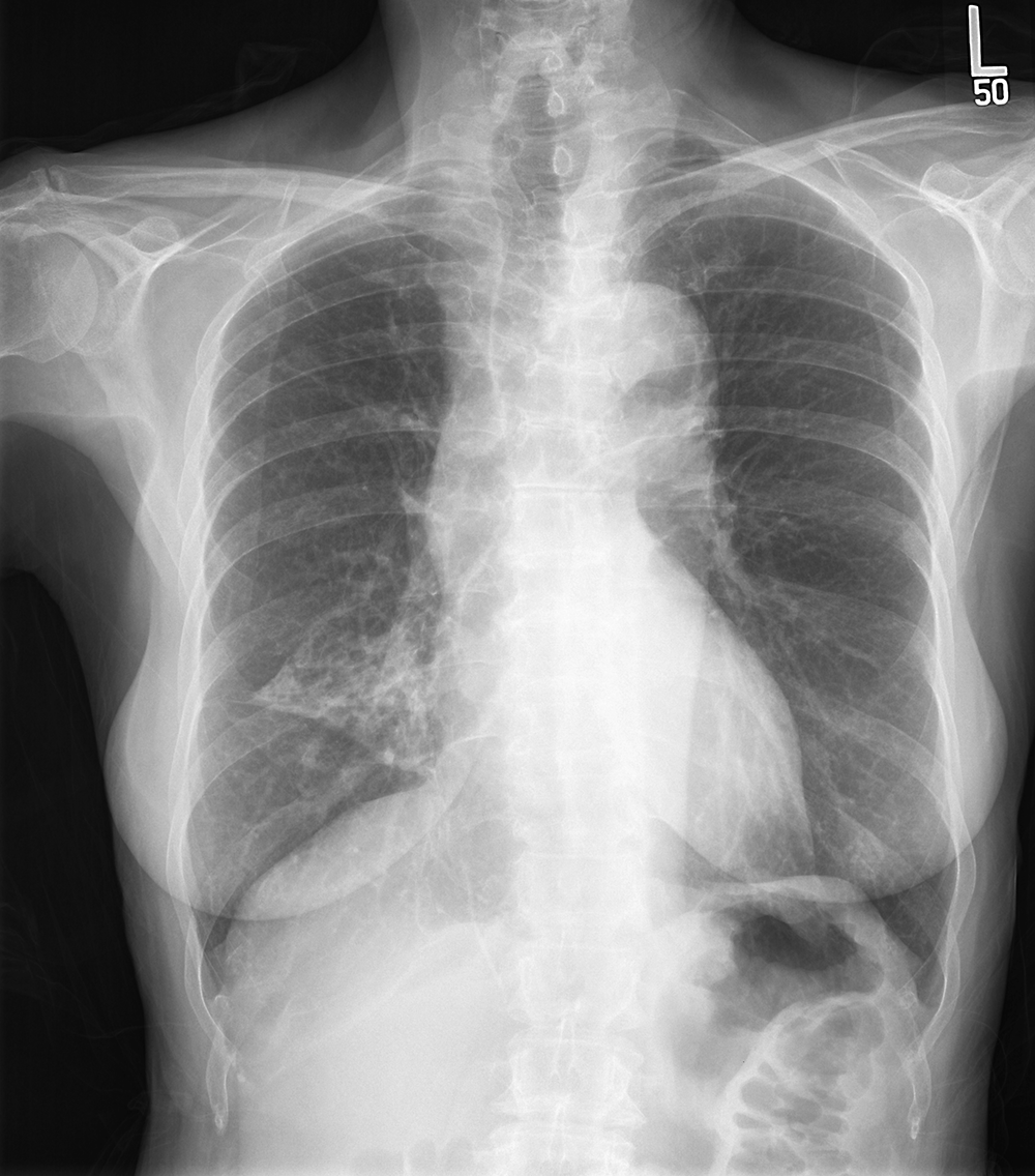
Case 3 concerns the way that problems with the airways appear on imaging. This 75 year old female developed hemoptysis. She has no smoking history. She has no fever or other symptoms other than mild recent fatigue.
Question 1:
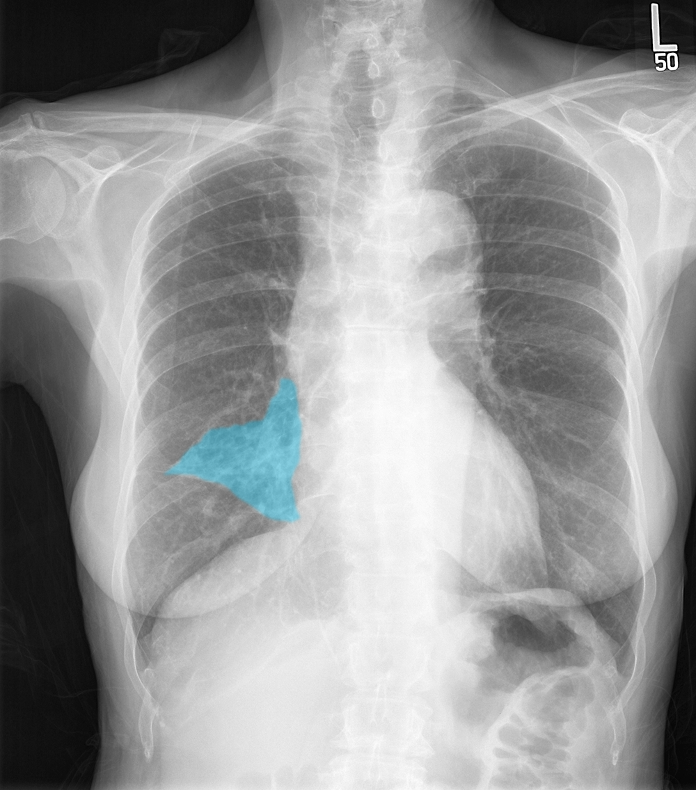
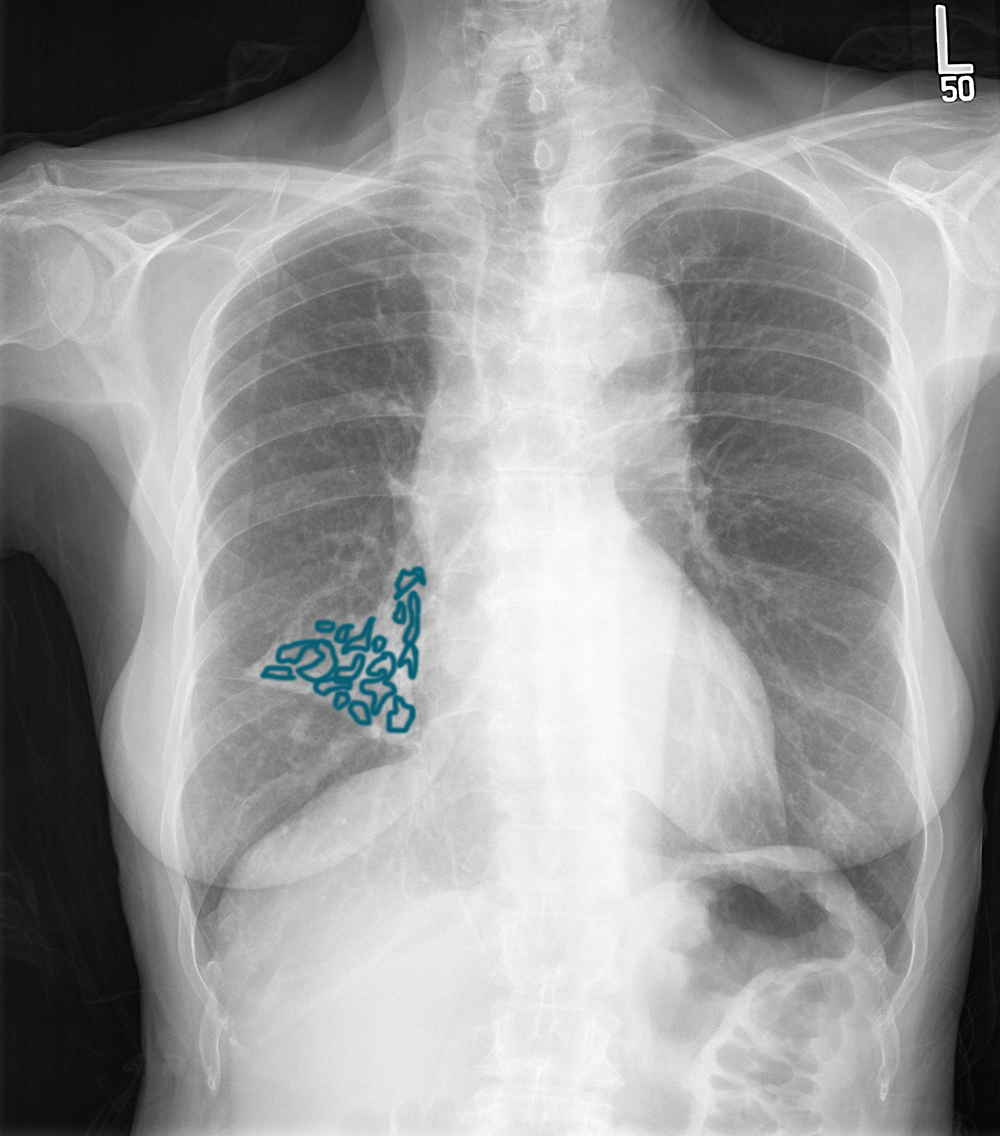
What are some descriptive terms you might use for the frontal chest radiograph? What do you call dilated bronchi? Given the location of the abnormality on this view, where would you expect to see it on a lateral view?
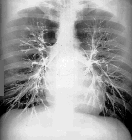
There is an area of abnormal opacity in the mid-right lung. It is overall somewhat triangular in shape, possibly representing a partly collapsed right middle lobe. Within the area, there are rounded lucencies, some of which appear slightly tubular. Since the structures appear somewhat tubular and air-filled, this should suggest the bronchial tree. When bronchi are dilated, this is called 'bronchiectasis'. As noted above, the location of the abnormality suggests involvement of the right middle lobe. This would appear anteriorly, overlying the heart, on a lateral view. The mystery study below (done on a different patient) is called a bronchogram. It is no longer done, but was a way to see the bronchi better in the days before CT. The patient inhaled an oily contrast material that coated the bronchi.
Case 3
This is the next study that was done on this patient.
Question 2:
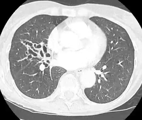
What are pertinent technical parameters to note on this image? What else is abnormal about the right middle lobe besides the dilated bronchi that fill it?
This is an axial CT scan of the chest at the level of the middle of the heart. The image is displayed in a lung window, which makes it a bit more difficult to tell if IV contrast was given. The other thing that is abnormal about the right middle lobe is that the overall volume of the lobe is quite low, indicating scarring and loss of normal lung parenchyma. With this much bronchiectasis, that is not surprising, as this is often a chronic inflammatory process, and when bronchi become this abnormal, the lung they supply is also damaged due to lack of normal ciliary motility to facilitate drainage of secretions.
