


Case 2
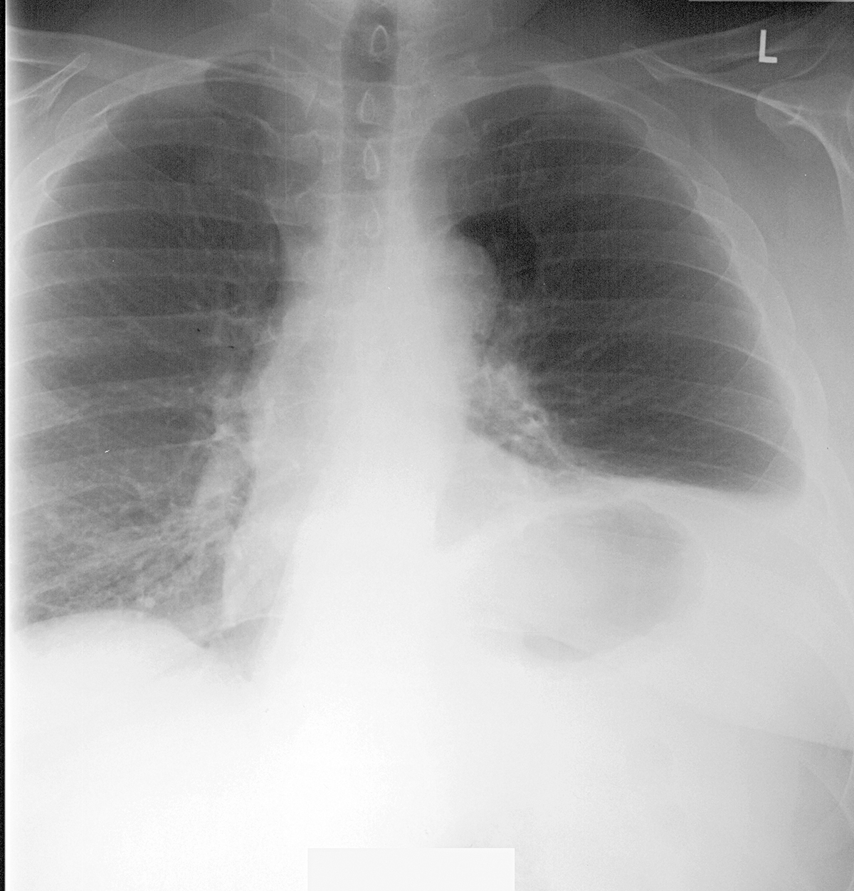
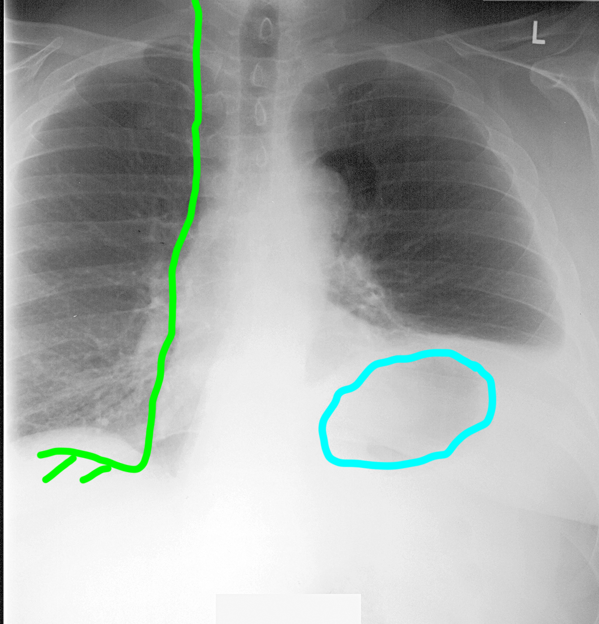
This is imaging from a 55 year old male patient with chronic shortness of breath. He has a past history of biopsy of some nodes in his mediastinum, but he does not recall what the procedure was called. He is a poor historian. He has a small scar near the left anterior 1st rib.
Question 1:
a) What is this image? Be specific.
This is a PA radiograph of the chest.
b) What two things are the most abnormal on this image?
There is a round lucency in the region of the aortic arch, and the left hemidiaphragm is markedly elevated.
Try to trace the expected course of the right phrenic nerve on this image.
Case 2
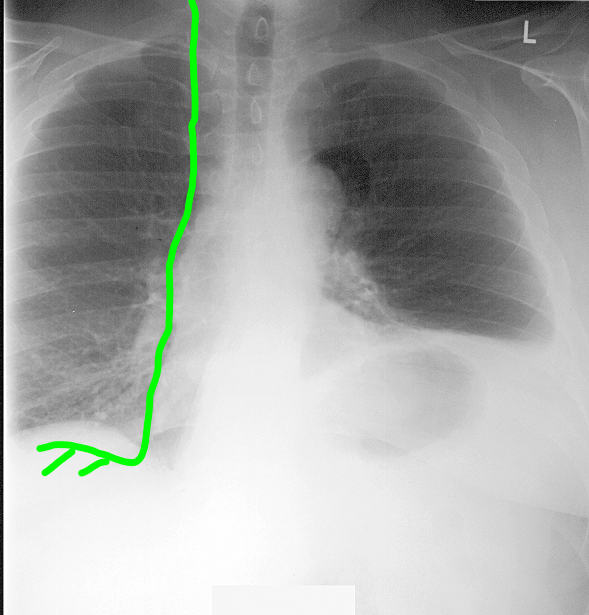
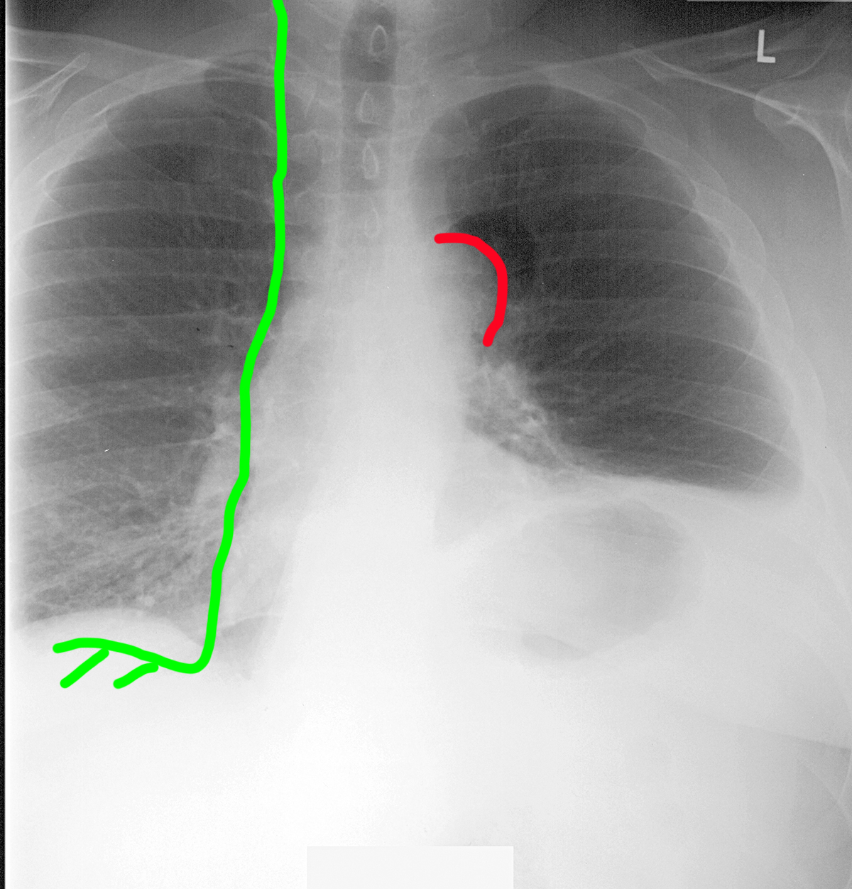
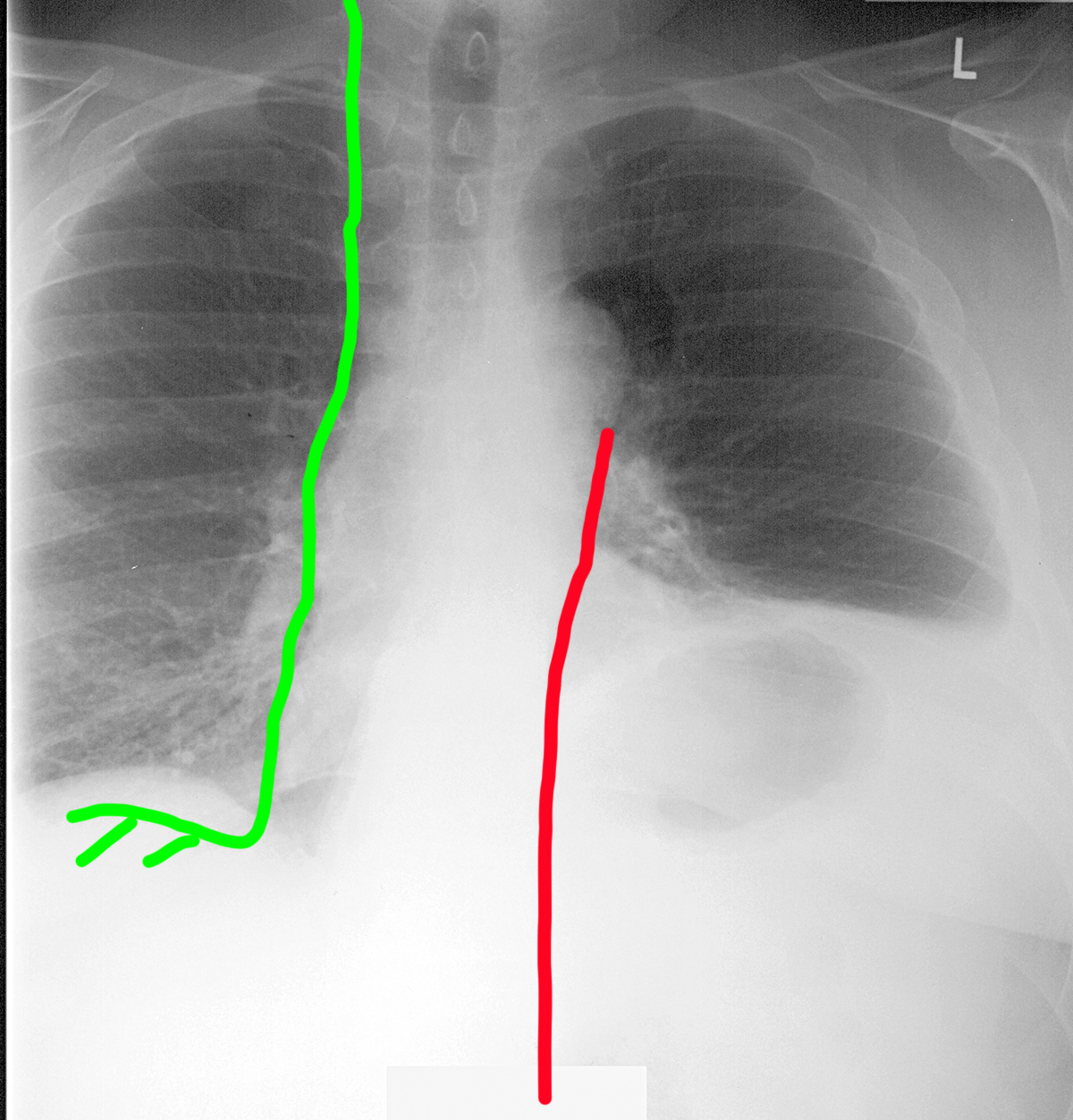
This is the same patient's x-ray showing the expected course of the right phrenic nerve in green. Try to identify the listed structures below before clicking the links.
Question 2:
a) Does the phrenic nerve pass anterior or posterior to the hilum of the lung?
The phrenic nerve passes ANTERIOR to the hilum. The vagus nerve passes posterior to the hilum, giving branches to the esophagus as it moves toward the abdomen.
b) What would happen to the position of the diaphragm if the phrenic nerve were damaged?
When the phrenic nerve is damaged, the diaphragm will move UPWARD, due to pressure from the abdominal contents and loss of the normal muscle tension that pulls the diaphragm downward during inspiration.
Case 2
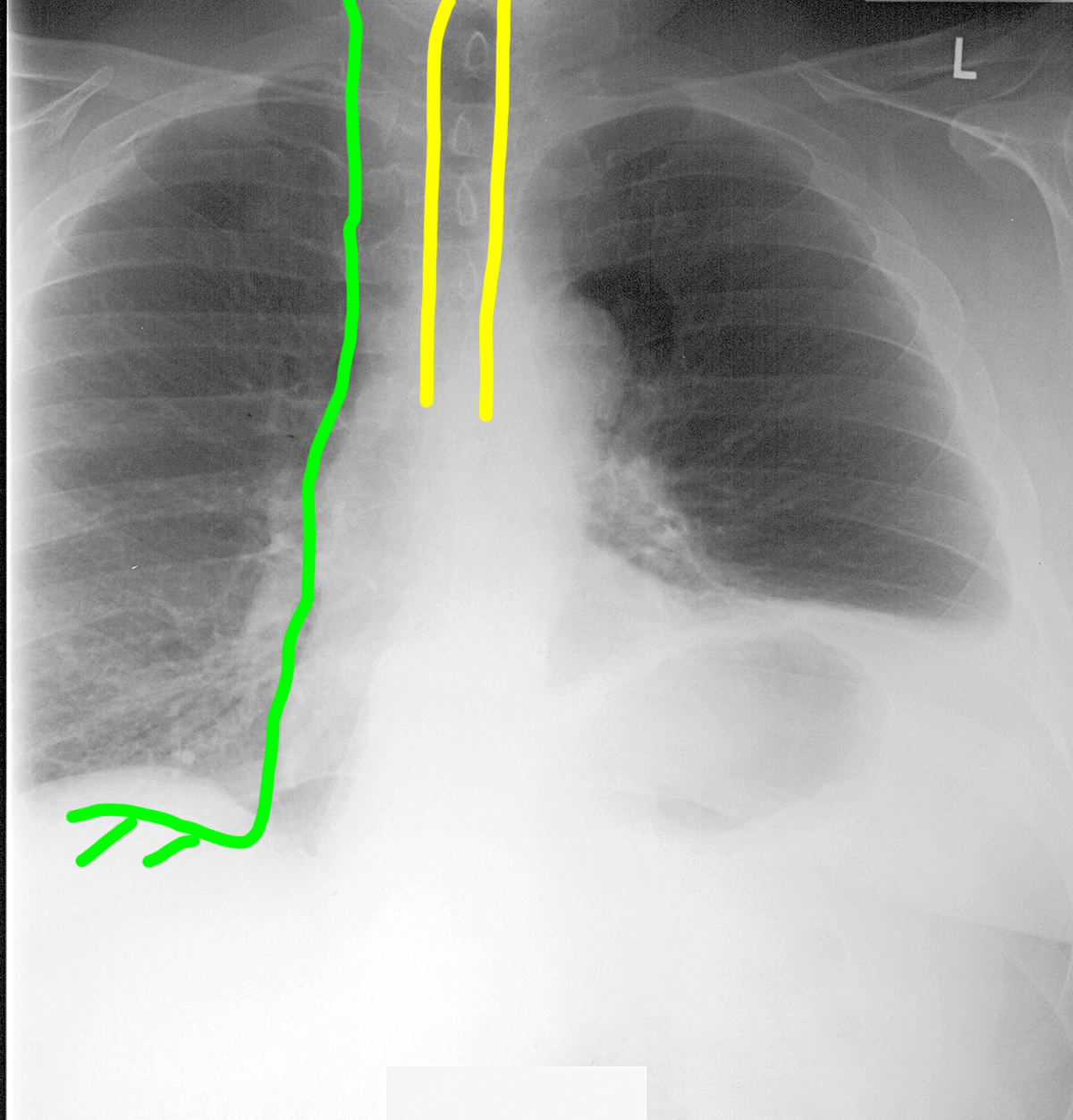
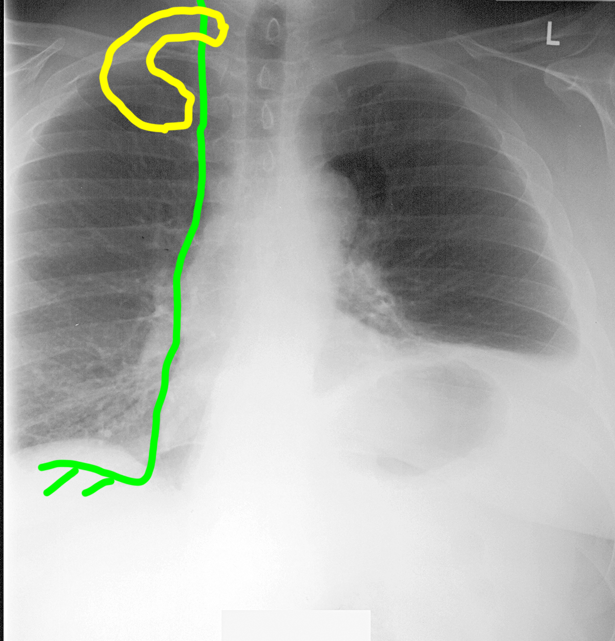
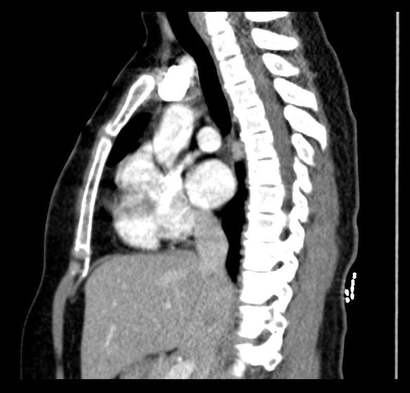
This is an image of a normal patient. There are three common ways to access nodes in the mediastinum for biopsy: via bronchoscopy (a tube placed into the tracheobronchial tree, with biopsy through the bronchial wall) and via mediastinoscopy (a tube passing into the mediastinum via a chest wall incision). Mediastinoscopy can be either CERVICAL (through an incision just superior to the manubrium) or ANTERIOR (through an incision lateral to the upper sternum, between upper anterior ribs).
Question 3:
a) What is this study? Be specific!
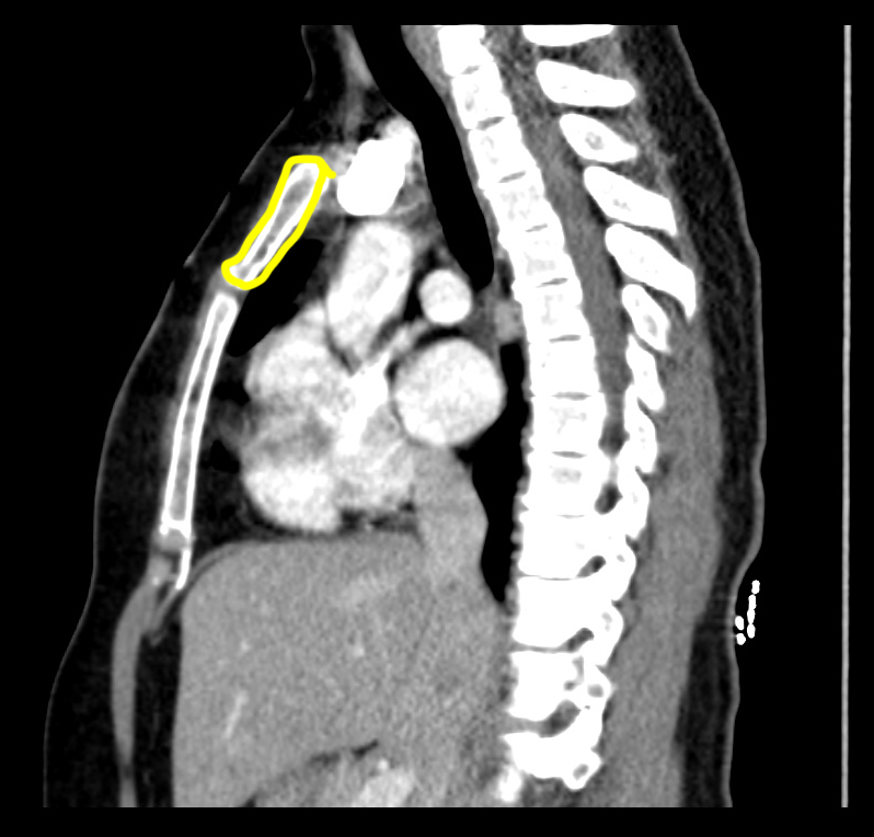
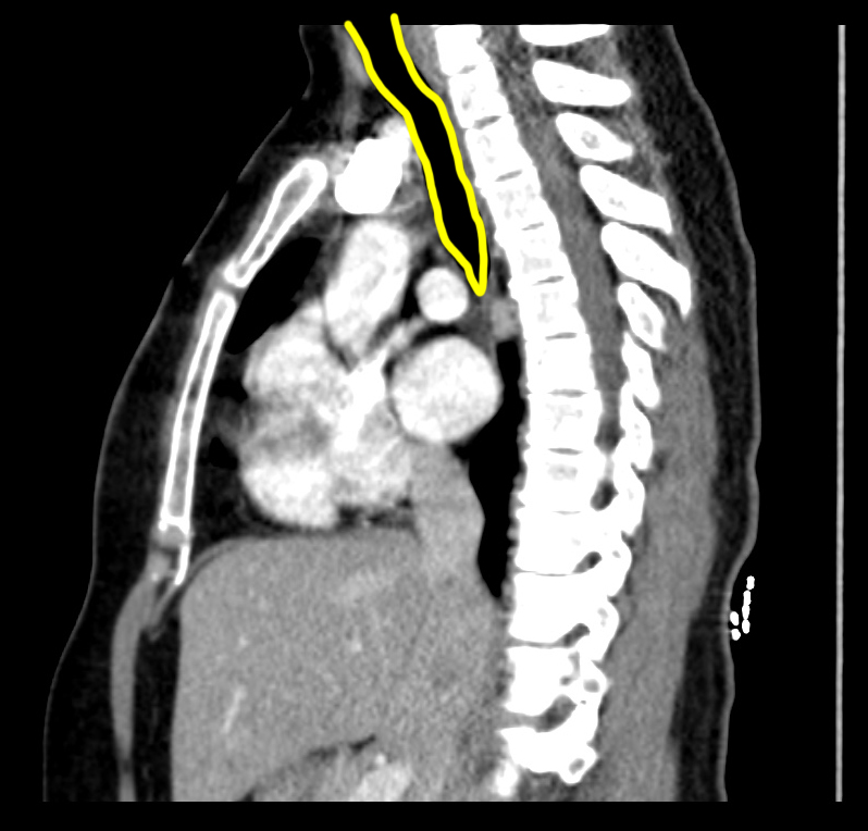
This is a CT scan of the thorax, reconstructed in the sagittal plane, and displayed with soft tissue windows.
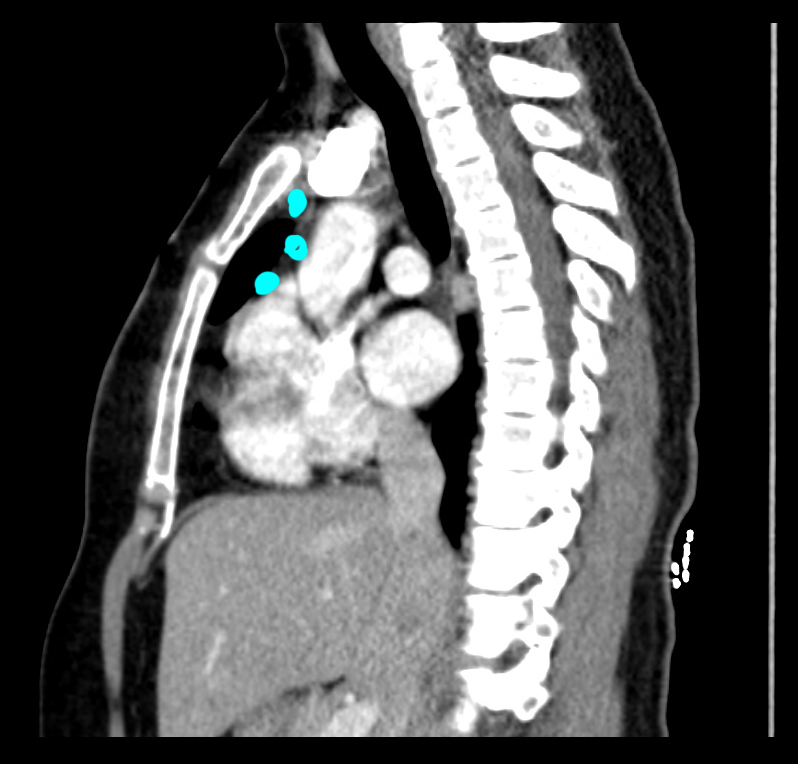
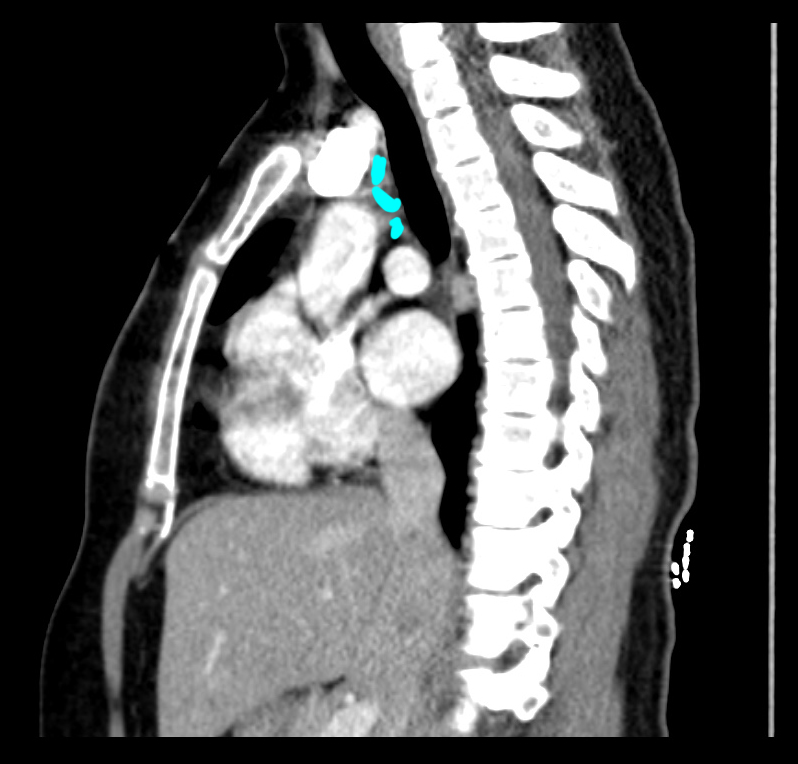
b) Identify the trachea and the manubrium on the image before clicking the links below. Which of the three sets of nodes shown below would best be accessed via anterior mediastinoscopy?
Node group 1 is most appropriate for anterior mediastinoscopy, where the incision leads the scope into the anterior mediastinal compartment. Node group 2 could be accessed via cervical mediastinoscopy or trans-bronchial biopsy. Node group 3 could only be reached with trans-bronchial biopsy because cervical mediastinoscopy runs along the anterior or lateral margins of the trachea.
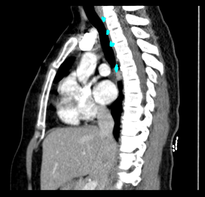
Case 2
This is a set of images of another study on the original patient. Links below provide a review of the thoracic spinal components visible on these images.
Question 4:
a) What is this study? Be specific!
This is a set of CT images of the upper chest, displayed with lung windows in the axial plane.
b) Do these images show a reason for the lucency seen in the region of the aortic arch?
YES! There is lung herniating out through the chest wall in this region, which is exactly where a mediastinoscope is often inserted, sometimes after resection of part of the rib and/or costal cartilage.


Case 2
This movie shows the approach for cervical mediastinoscopy, which is done through a small incision in the region of the top of the manubrium. The mediastinoscope is then passed along the anterior wall of the trachea.
Question 5:
What other approach could be used to reach these nodes?
Nodes can also be biopsied via bronchoscopy--a tube introduced into the tracheobronchial tree, with biopsy through the wall (trans-bronchial biopsy). Identification of the nodes from within the trachea can be challenging.


