

Case 3
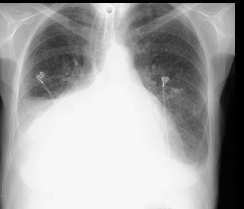
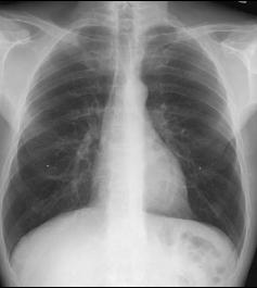
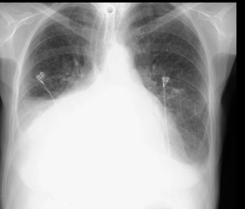
This image is from an 89 year old female with Alzheimers disease, coming in for a routine checkup. She is a poor historian and has a midline anterior chest scar from some sort of prior surgery, she is not sure what. She also has a tracheostomy tube, a tube in her nose, and is short of breath. A normal image from a different patient is available to you using the link below.
Question 1:
a) What is this study?
This is a chest radiograph. It is hard to tell if it is AP or PA without labels. Given the patient's status, AP is likely.
b) What do you think of her heart?
The right side of her heart is hard to see, but the left side seems to suggest that it is enlarged.
c) What else looks abnormal?
Both costophrenic angles appear hazy with a suggestion of a curved configuration (meniscus). This suggests the presence of pleural fluid.
Case 3
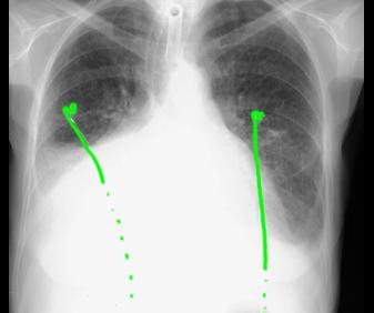
Here is our patient's radiograph again. Try to identify the support lines that are present before clicking on the links below.
Question 2:
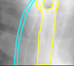
a) What is shown in the enlarged image in yellow?
The yellow outline shows the tracheostomy tube, placed in patients who require long term ventilatory support. The tube exits from the trachea through an opening in the anterior neck.
b) What is shown in the enlarged image in blue?
The blue outline shows the course of an NG tube. This is a plastic tube that is smaller in caliber than the tracheostomy tube (or an endotracheal tube), usually going from the nose into the stomach. This can be used to either drain fluid from the stomach in cases of obstruction, or to administer feeding support or medications directly into the stomach.
Case 3
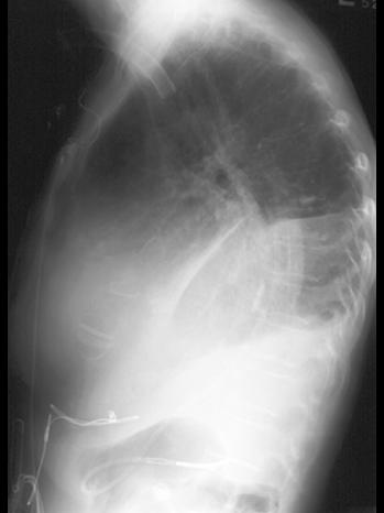
This is another image of our patient.
Question 3:
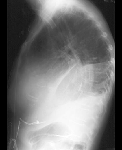
a) What is this view?
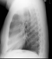
This is a lateral radiograph of the chest.
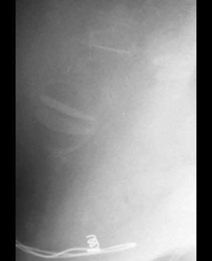
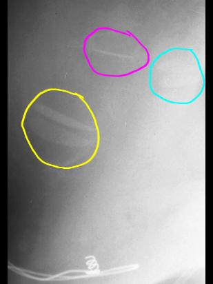
b) There are faint objects projecting over the heart that are seen best on the closeup view (see link below). What are they?
The objects appear too geometric in shape to be part of the body. Since they are overlapping the heart, and the patient has a midline anterior chest scar (suggesting prior cardiac surgery), these most likely represent artificial heart valves.
Case 3
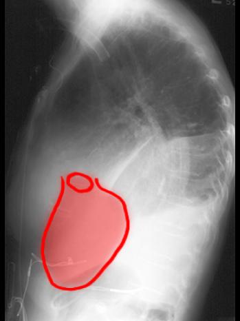
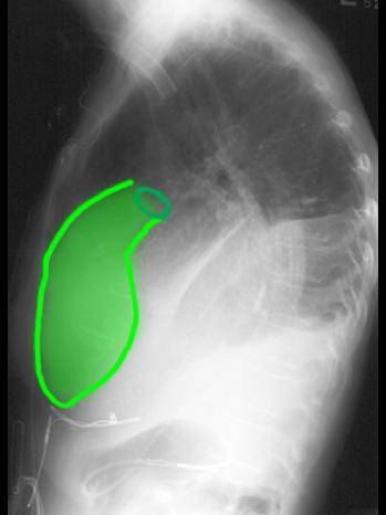
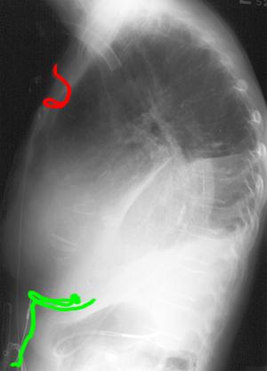
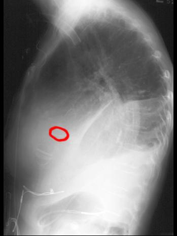
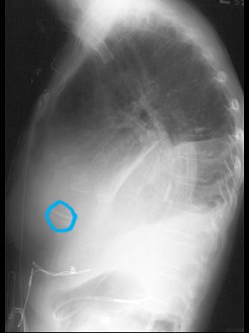
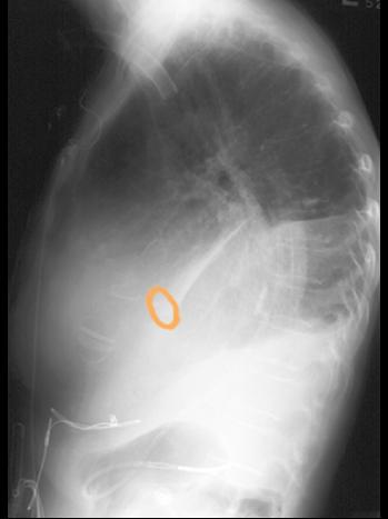
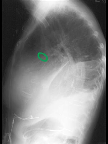
This is our patient's lateral radiograph again. There is a sternal wire outlined in red, and a pacer outlined in green.
Question 4:
a) The heart valves in this patient are not easy to see. What are they likely made of?
Many artificial valves have metal components, and these are usually easy to see on radiographs. These valves are likely made of plastic or of natural materials (such as porcine valve material) which are much closer to the density of the natural soft tissues of the heart, making them hard to see on a radiograph.
b) What other imaging modality would likely show the valves more clearly?
Either ultrasound or CT would likely show the valves more clearly. MRI could be considered, but it would be important to first be sure that the valves are MR-compatible before putting the patient into an MR scanner. Given the lack of adequate history for this patient, this seems like it would not be a good idea.
Case 3
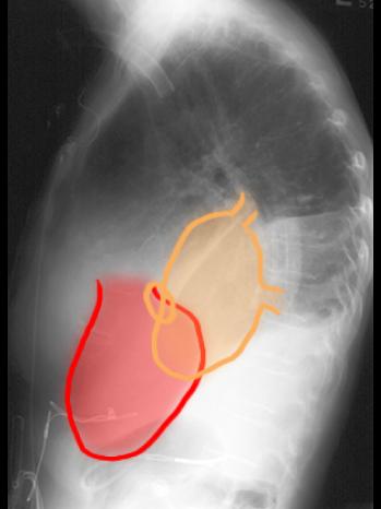
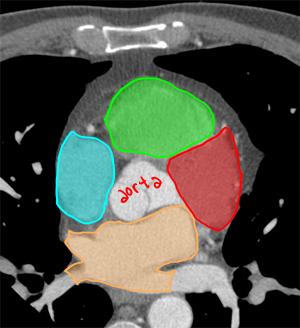
In order to figure out which valves have been replaced, it is helpful to review the orientation of chambers in space, best shown on cross-sectional imaging.
Question 5:
a) What type of study is this? Be specific.
This is an axial CT study, displayed with bone or IV contrast windows (optimized to show relatively dense structures), with IV contrast in the early arterial phase.
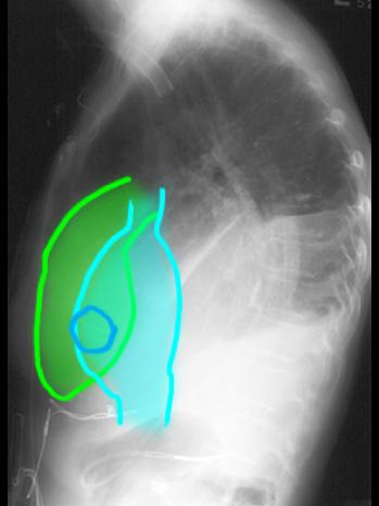
b) From the single orientation image (link below), decide which chamber is which.
Light blue is the right atrium. Green is the right ventricle. Red is the left ventricle. Orange is the left atrium.
Case 3
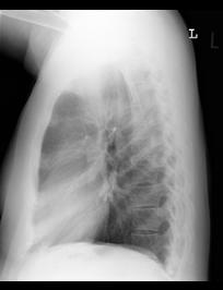
This is our patient's lateral chest radiograph again.
Question 6:
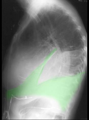
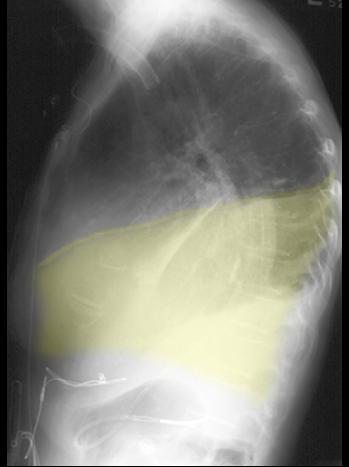
What do you think abnormalities 1 and 2 represent on the labels provided?
These are both pleural effusions. From the frontal view, we know that the right effusion was larger, so it is abnormality 2. The left effusion is abnormality 1.
Case 3
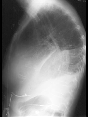
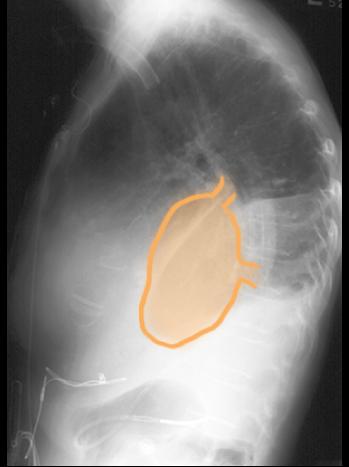
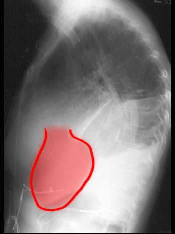
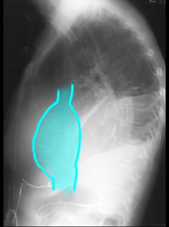
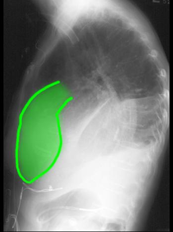
This is again our patient's lateral chest radiograph, with the valves and associated chambers indicated by labels below.
Question 7:
a) What valves are most commonly replaced?
Aortic and mitral are most common.
b) What valve is located most anteriorly in the heart?
The tricuspid valve is most anterior in location.
