

Case 3
This set of images is from a 63 year old man with sudden onset of severe abdominal pain, and how has developed thrombi in various sites within his body suggesting a hypercoagulable state.
Question 1:
a) What type of study is this? Be specific.
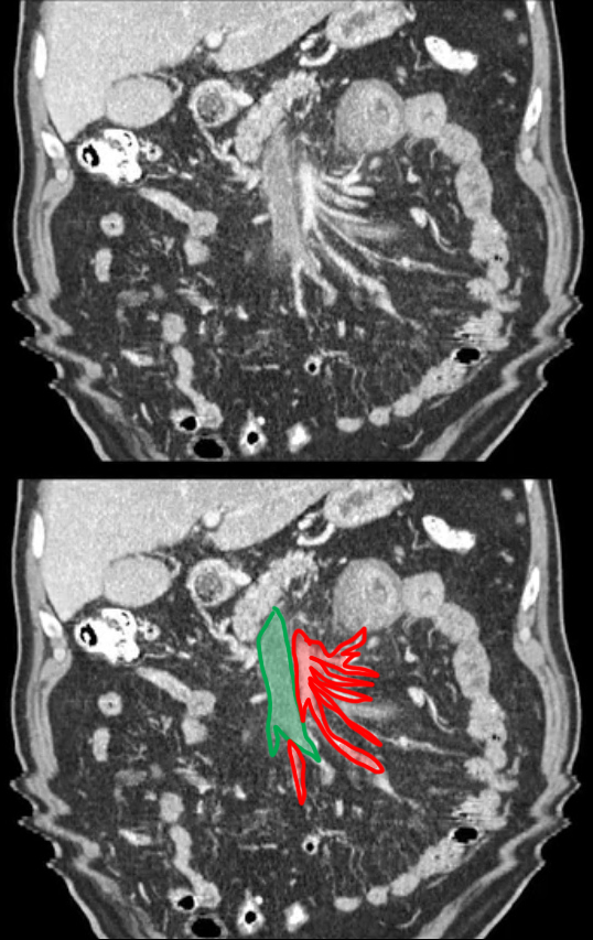
These are coronal reformatted CT images, displayed with soft tissue windows, with both oral and intravenous contrast present.
b) What is a possible explanation of the jagged appearance of the lower part of the images?
The patient may have coughed or started breathing quickly before the entire scan series was finished.
Case 3
This is the same coronal CT series, with three vascular structures labeled for you to identify.
Question 2:
a) What is labeled in red?
The red structure is the SMA. You can identify it because it starts out in the midline coming off the aorta, then turns inferiorly, giving off many branches.
b) What is labeled in green?
The green structure is the SMV. It runs in parallel with the SMA, but is normally slightly larger in diameter. This SMV is abnormally large.
c) Whatis labeled in light blue
The portal vein is labeled in light blue, entering the portal hepatis at the bottom of the liver. It is formed by the junction of the SMV and the splenic vein, which is not shown in these slices as it lies further posterior in position.
Further Explanation:
Case 3
This is a selected slice from the CT series previously shown with unlabeled and labeled images.
Question 3:
a) How can you tell that the red structure is NOT the aorta and the green structure is NOT the IVC?
The slice is too far anterior within the abdomen, with only bowel loops visible along with the liver. The more posterior structures such as the spleen and kidneys, are not visible. The aorta lies in the same posterior plane as other retroperitoneal structures. Also, the aorta does not have this many branches coming off laterally.
b) Does the SMV look normal?
No. The SMV should be only slightly larger in diameter than the SMA, so this vessel is too large. Also the density (attenuation) in the lumen of the SMV seems too low. It should be more similar in density to the SMA. This suggests that something else is in the lumen, like thrombus.
Case 3
This set of images is from a different patient. Try to find the SMA and SMV as well as the aorta and IVC.
Question 4:
a) How does the density of the SMA and SMV differ in this example from our previous patient?
In this patient, the density in the lumen of SMA and SMV is much more similar. The SMV is slightly larger than the SMA, but not as much as in our previous patient. Incidentally, this patient has mural (wall) calcification in the SMA consistent with atherosclerosis.
b) What other organs are visible on slices with the aorta and IVC visible?
Pancreas, kidneys, upper portions of the ascending and descending colon and iliac crests, all posterior structures. This patient has had a splenectomy, so no spleen is visible.
Case 3
This is a single labeled image from the previous case. Try to identify the outlined structures.
Question 5:
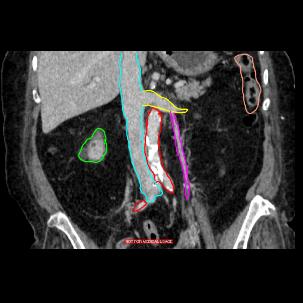
a) What is outlined in green?
The anterior portion of the right kidney.
b) What is outlined in light blue?
The IVC is outlined in light blue, passing through the posterior portion of the liver.
c) What is outlined in red?
The aorta and part of the right common iliac artery, with extensive atherosclerotic calcification, is outlined in red.
d) What is outlined in purple?
The left gonadal vein, draining into the left renal vein (outlined in yellow)
e) What is outlined in orange?
The upper portion of the descending colon, near the splenic flexure.
Case 3
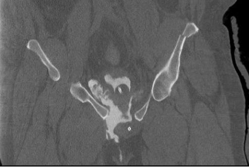
This is an image of a different patient with very similar symptoms--acute onset of severe diffuse abdominal pain.
Question 6:
a) What kind of study is this?
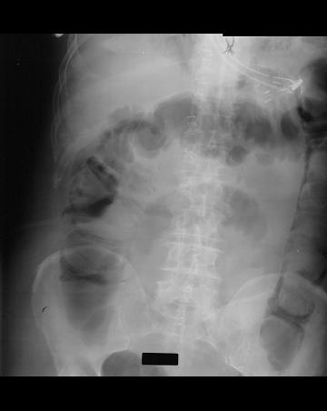
This is a KUB, presumably in supine position.
b) Where is most of the gas on this image?
Most of the gas is in the colon, but there is a single small bowel loop more centrally that looks dilated.
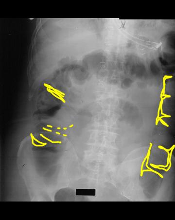
What do you think the mystery label below indicates?
Case 3
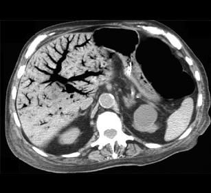
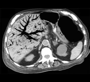
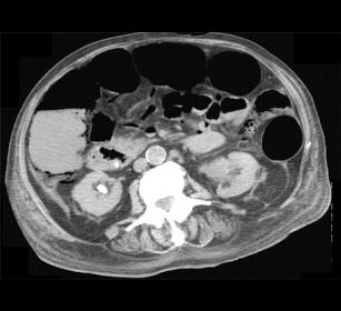
The mystery on the prior image showed gas that was in small streaks or pockets in the periphery of the colon, most consistent with gas in the wall of the bowel (pneumatosis intestinalis). This is often a sign of severe infection or infarction of bowel, with breakdown of the wall. Gas, which is normally present in the lumen of the colon can then move into and through the wall.
Question 7:
a) What do you notice on the CT slice presented that appears unusual?
There is a branching black pattern within the liver, indicating gas in the portal venous system.
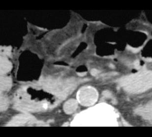
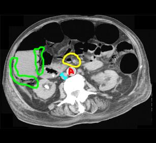
b) On the lower CT slice (see link below), what do you think of the right colon?
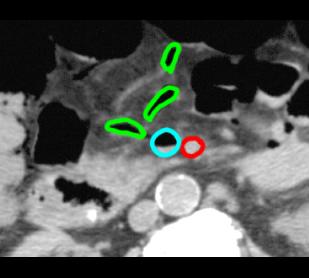
There is gas in the wall of the right colon, indicated in green in the labeled image (link below).
c) What is circled in yellow on the lower CT slice with labels?
The yellow circle indicates the location of the SMV and SMA. The SMV contains gas.
Case 3
This is the upper CT slice through the liver on the same patient.
Question 8:
a) How can you connect the KUB finding of pneumatosis intestinalis with the appearance of the liver on this image?
When gas goes from the lumen into the wall of the colon, it can make its way into the portal vessels that drain the GI tract. This explains the gas present in the SMV. Once in the SMV, the gas can make its way into the portal vein and then into the liver. Portal venous gas is a very bad sign, usually indicating a severe problem with the GI tract and breakdown of the walls of the viscera.
b) What are the outlined structures on the closeup CT image with labels?
The light blue circle is around the SMV, with has an air-fluid level, meaning that it is partly filled with blood with air floating to the top. The SMA is outlined in red and appears normal. The green outlines are various other portal tributaries making their way to the SMV, and filled with gas.
