

Case 3
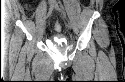
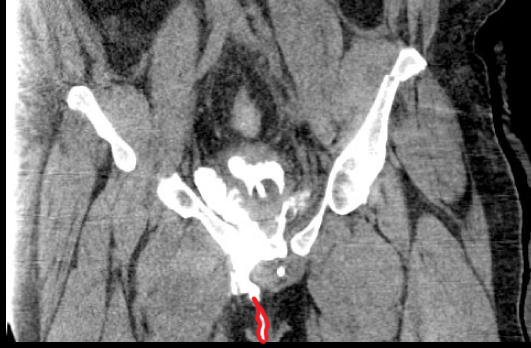
This is imaging of a 23 year old male who was involved in a high speed motor vehicle collision, with severe pelvis and abdominal pain.
Question 1:
What kind of study is this? Be specific.
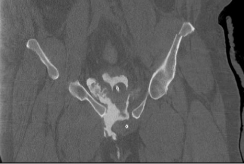
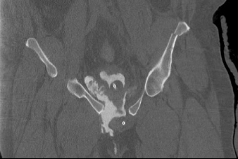
This is a pelvic CT scan, reconstructed in the coronal plane and displayed with bone windows. No oral or intravenous contrast is present but there is contrast in the bladder.
Case 3
This is a selected image from the patient's CT
Question 2:
a) How do you know that this is a bone window?
You can tell that this is a bone window because of the internal detail you can see within each bone.
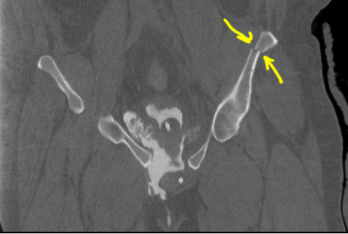
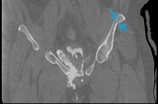
b) Is there a bony abnormality?
Yes, there is a fracture of the left iliac bone with slight displacement.
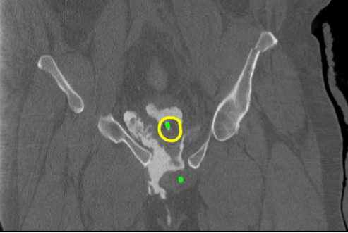
c) What is the structure indicated in yellow and green below?
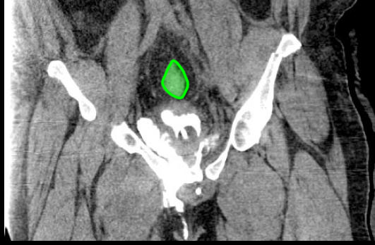
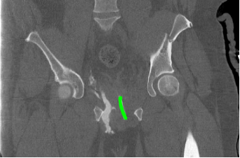
This is a bladder catheter (green) with a balloon blown up in the bladder (yellow).
Case 3
This is a selected image from the previous CT series.
Question 3:
a) How did the contrast get into the bladder?
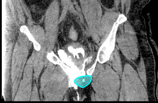
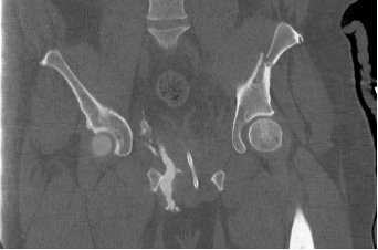
Since there is no contrast in the vessels, it was not excreted from the kidneys into the bladder. It was injected through the bladder catheter indicated on the previous page. The rest of the catheter is outlined below on the second slice
b) What else is wrong with the pubic bones on this image?
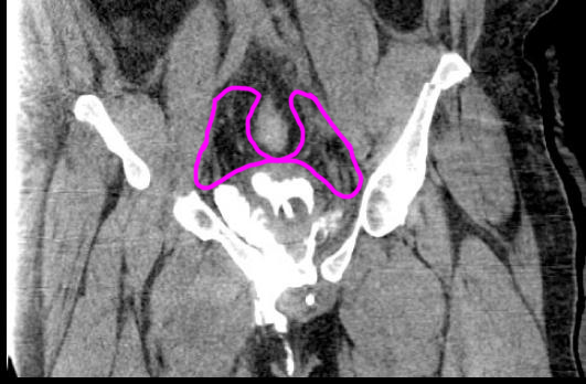
This image shows the flat surfaces on the medial sides of the pubic bones, which should come together to form the pubic symphysis. They are much too far apart, indicating that the symphysis has been fractured apart and separated by the trauma.
Case 3
This study is from a different patient, and is a chance for you to practice identifying different CT windows.
Question 4:
a) What is the imaging plane for these studies?
These are CT studies reconstructed in the coronal plane.
b) Was IV contrast given?
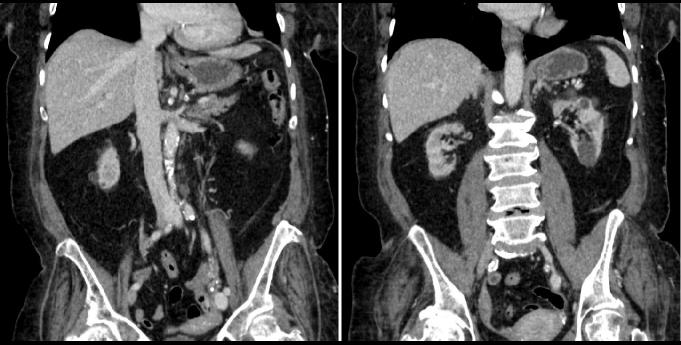
Yes, the aorta is brighter than muscle, as is the liver, spleen and kidneys.
c) What window was used for each set of images?
The initial set of images is displayed using a bone window. The calcifications along the aorta are particularly well shown as are bones. The set below 'other window 1' is shown in lung windows. The set below 'other window 2' is shown in soft tissue windows.
Case 3
These are the soft tissue windows from the same case.
Question 5:
a) Did the patient need extra radiation dose to perform the bone, lung and soft tissue windows for this study?
No. The various different windows can be adjusted after the study is completed and do not add to patient radiation dose.
b) Did the patient need extra radiation dose to reconstruct the images in the coronal plane?
No. The original data was collected in the axial plane, but different planes can be reconstructed after the study is completed and do not add to patient radiation dose.
c) If the patient had non-contrast scans before the IV contrast was given, did the patient receive extra radiation dose?
Yes. Each time the patient is put through the scanner another time this does increase the radiation dose. If the entire body was scanned before and after IV contrast, this would double the radiation dose.
Case 3
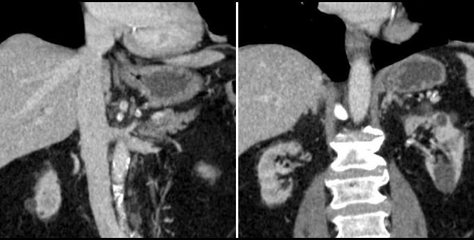
These are two selected images focusing on the kidneys in this same patient.
Question 6:
a) What tissues do you look at to identify a soft tissue window?
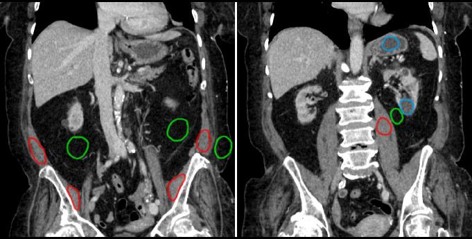
If you click on 'densities' below, you will see that muscle (red) and fat (green) are very different in shades of grey. This tells you that this is a soft tissue window, since it lets you see soft tissue structures outlined clearly by much darker fat.
b) What kind of material is in between fat and muscle in density?
Water is in between muscle and fat in density, and is circled in blue on the 'densities' image. There is water in the stomach as well as in multiple rounded structures in the kidneys. If we were farther posterior in the body, we might see water in the cerebrospinal fluid. If we were farther anterior in the body, we might see water in the gallbladder or urinary bladder.
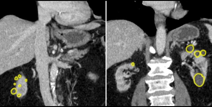
c) What is the mystery, indicated below?
The rounded water density structures in the kidneys are cysts. These are very common and usually of no clinical importance unless they are so numerous and large that they replace the renal parenchyma and impair renal function. The cysts in this case are not clinically important.
Case 3
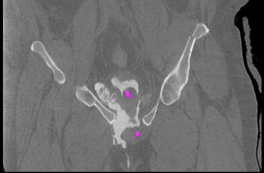
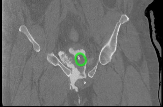
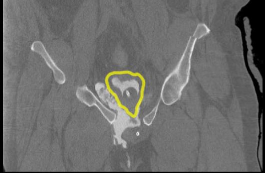
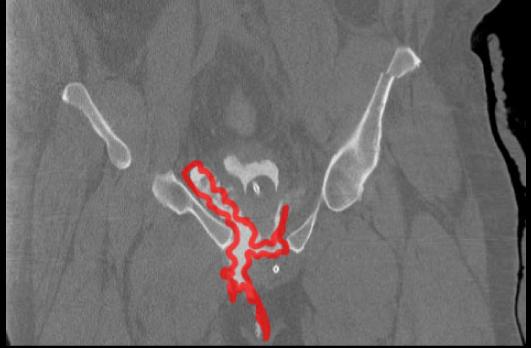
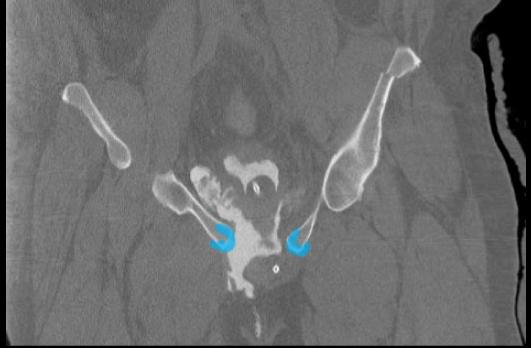
This is one of the CT images from our original case, the patient who had significant trauma in a motor vehicle collision, and severe pelvic and abdominal pain. Try to identify the structures indicated below before clicking the links.
Question 7:
Into what space is contrast leaking in this patient?
The bladder is covered on its superior surface with peritoneum, but the remainder of the organ is extraperitoneal. Bladder rupture can occur at the uppermost portion, in which case contrast would leak into the peritoneal space. In this patient, the rupture was in the lower part of the bladder, and is leaking down into the extraperitoneal space.
Case 3
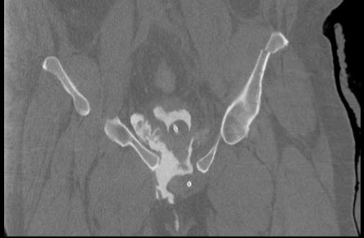
This is another image of the same patient's study. Try to identify the listed structures or findings before clicking on the links.
Question 8:
a) What is this window?
This is a soft tissue window. Notice that the bony detail and the exact borders of the extravasated contrast are not as clearly seen.
b) Where would the contrast collect in an intraperitoneal bladder rupture?
Intraperitoneal bladder ruptures are much less common than extraperitoneal. In intraperitoneal rupture, the contrast would be emerging from the superior surface of the bladder, and would surround structures like the sigmoid and other adjacent bowel loops. It would not be found inferior to the bladder or extending down to the perineum or scrotum, as it is doing in this case.
