

Abdomen Pelvis Case 2
This 51 year old patient had decreased appetite and increased abdominal girth.
Question 1:
What potential abnormalities might be missed on a study with these technical parameters?
This is an axial abdominal CT scan with IV but no oral contrast (patient had nausea and could not drink the contrast), displayed with soft tissue windows.Subtle bony or lung findings might be missed on this study. Problems in the GI tract might also be harder to identify without GI contrast present. Some abnormalities are easier to assess with other imaging planes, such as sagittal or coronal.
Look at the other portions of the study provided below and decide if they show any additional findings.
Abdomen Pelvis Case 2
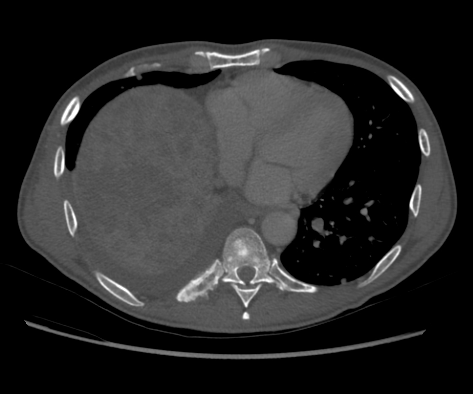
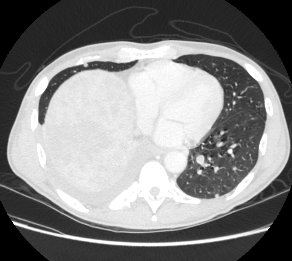
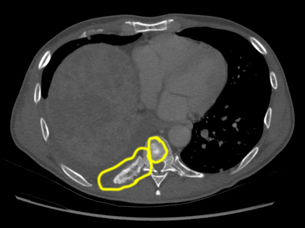
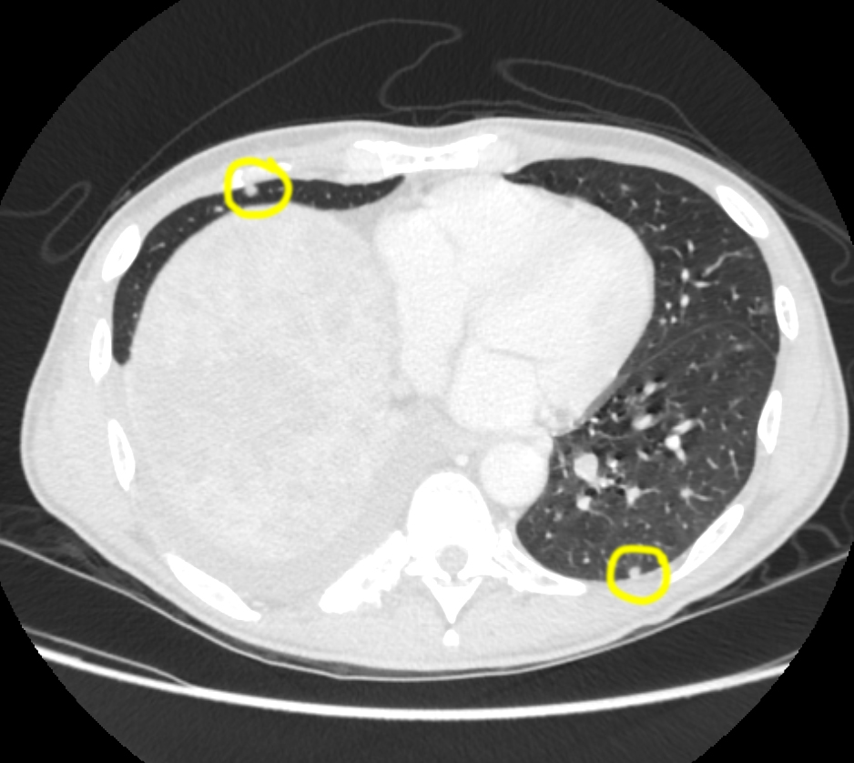
The coronal images (shown again here) demonstrate the degree of liver enlargement more clearly than the axial images. The bone window images show an unusual appearance of one of the posterior right ribs and one of the vertebral bodies that could represent tumor spread. The lung window images show a few tiny rounded densities (nodules) that might represent spread of the patient's tumor. Both abnormalities are circled below.
Question 2:
a) Does adding other windows and other imaging planes change the radiation dose for the study?
No. These changes can be made after the patient leaves the department and have no impact on how long the study takes or what the radiation dose is.
b) There is an abnormality of the peritoneal cavity on these images. What density is the abnormality?
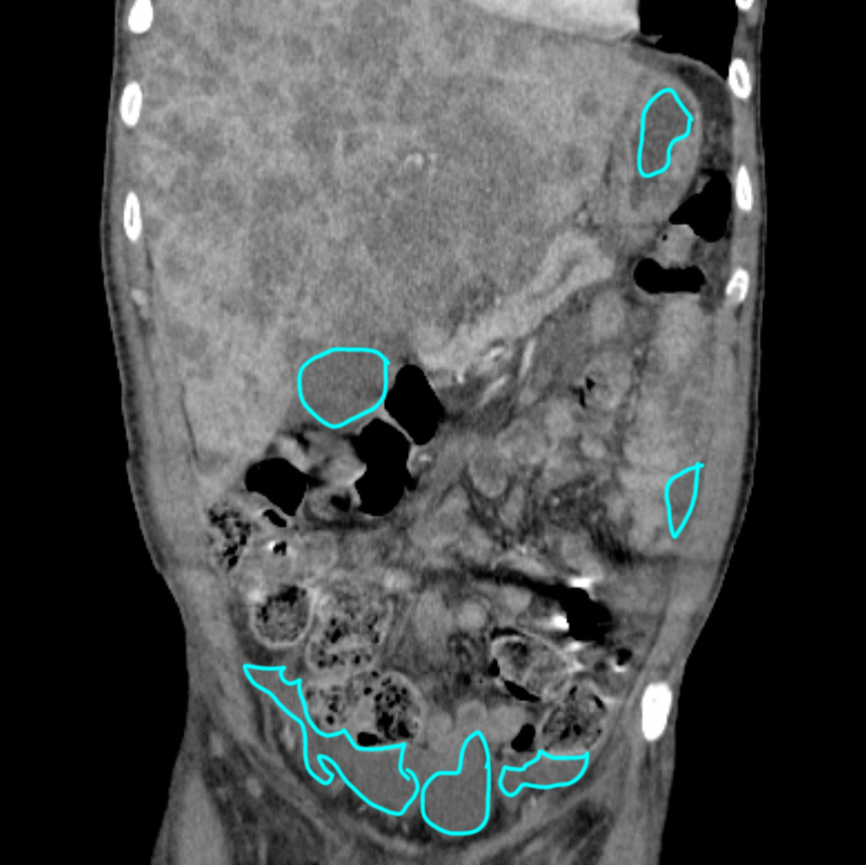
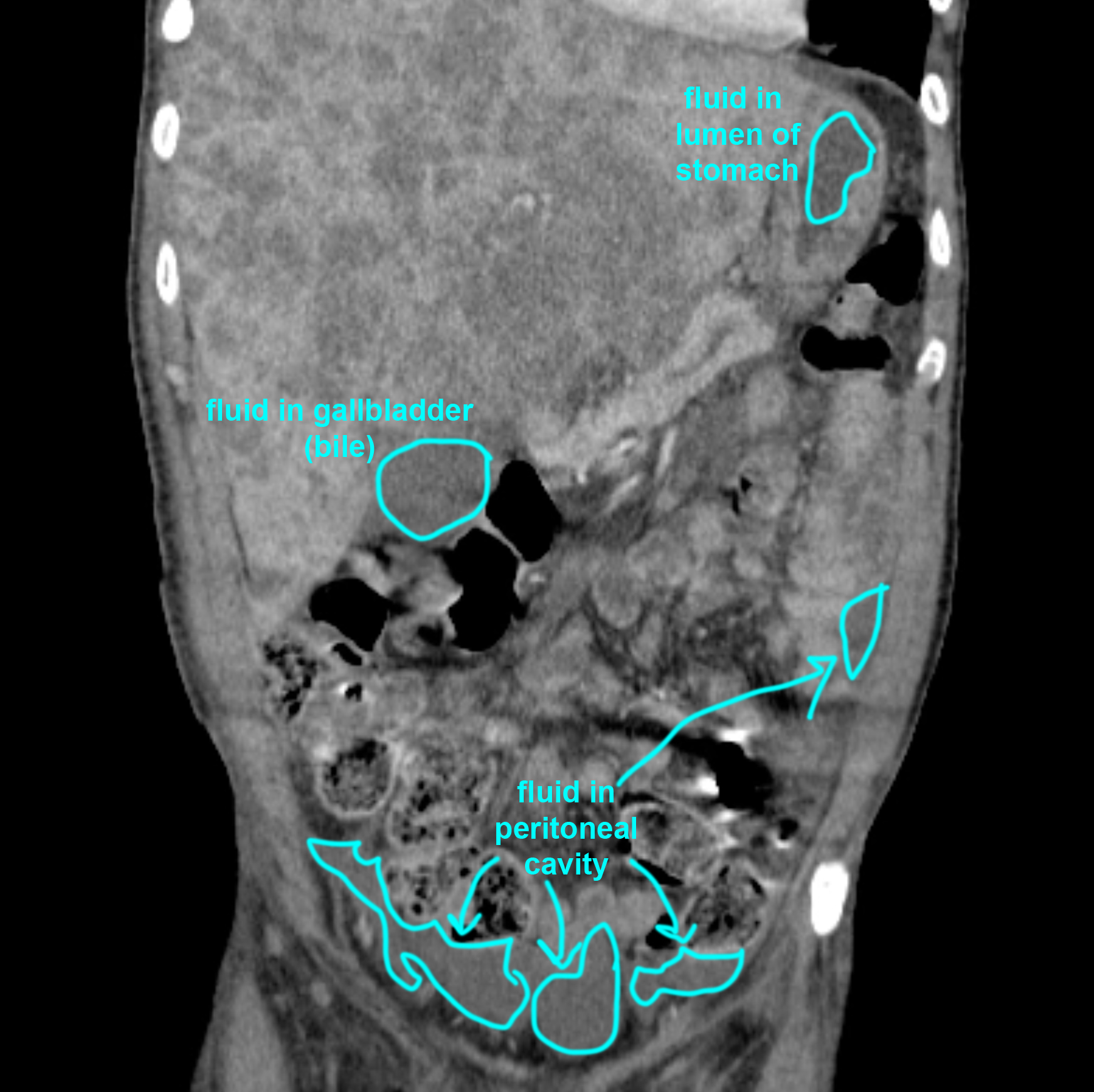
The peritoneal cavity is usually too thin to be visible on CT, but when it is filled with material as in this case, it becomes visible. The density of the material in the peritoneal cavity is water density. Click on the outlines below and try to identify the location of each area of water density before checking the labeled image.
Abdomen Pelvis Case 2
The liver in this patient is quite enlarged and abnormal in appearance, with many round low density lesions consistent with tumor deposits. For the rest of the discussion of this case, we will ignore the liver and concentrate on the rest of the abdominal organs.
Question 3:
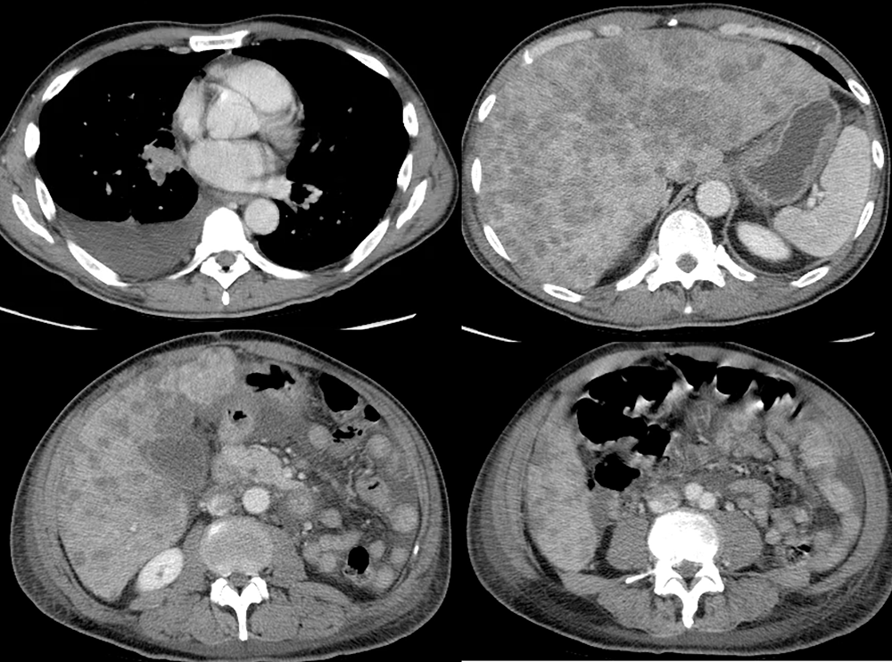
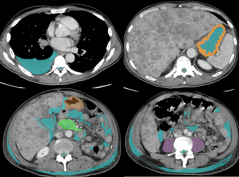
On this set of four images, where is fluid density evident, including normal and abnormal locations. Recall that fluid is darker grey than muscle, but not as dark as fat.
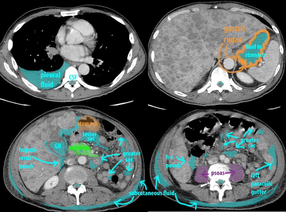
Normal sites of fluid density include the cerebrospinal fluid surrounding the spinal cord, fluid in the lumen of the stomach, and fluid in the gallbladder (in the form of bile). Abnormal locations of fluid in these images include fluid in the right pleural space, fluid in the peritoneal cavity including the lesser sac, hepatorenal pouch (Morrison's pouch), in between bowel loops in the greater sac, and the left paracolic gutter. Try to identify these locations on your own before clicking on the link below to display labels.
Abdomen Pelvis Case 2
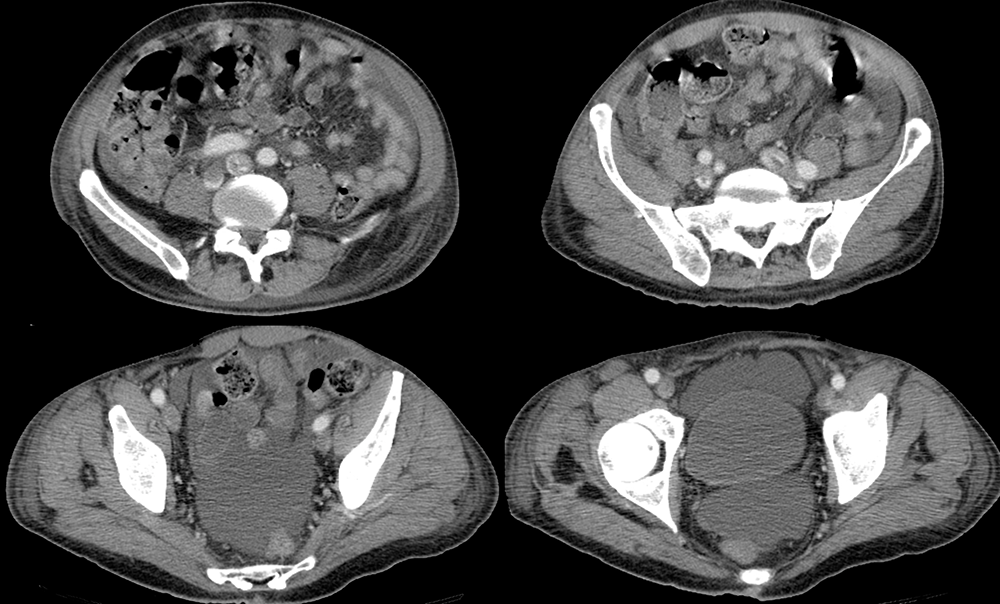
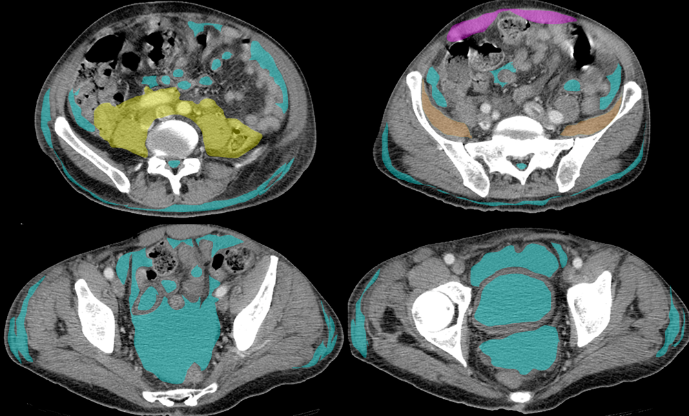
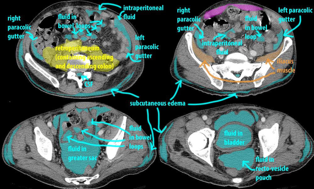
These four images are from the same patient, lower in the abdomen. Again, ignoring the very abnormal liver, try to identify where fluid is present before clicking on the links below.
Question 4:
a) Where do the lesser and great sacs connect?
At the porta hepatis (foramen of Winslow). This is not an easy location to visualize on axial CT images, but would be near the gallbladder fossa.
b) How can you tell if fluid on an abdominal CT image is inside vs outside of a bowel loop?
Fluid in a bowel loop (which is normal in the stomach, small intestine, and right side of the colon) tends to have a rounded, oval, or tubular shape on a CT slice. Fluid that is outside the bowel loops (which is not normal, but shows you where the peritoneal cavity is located) tends to be irregular in shape, conforming to the outer walls of many adjacent bowel loops and organs. These images contain many examples of each for you to review.
c) How is the patient positioned for an abdominal CT scan and where will peritoneal fluid tend to accumulate in this position?
Patients are almost always supine for abdominal CT scans, although other positions are possible in special circumstances (prone, decubitus, sometimes determined by patient comfort). In the supine position, fluid tends to fall to the most posterior location, which is the recto-vesicle pouch. Other relative low points may include the lesser sac, the parabolic gutters, and the hepato-renal pouch. Try to find these locations on your own before clicking the labels link below.
d) What portions of the colon are retroperitoneal in location?
Most of the ascending and descending colon are retroperitoneal in location, moving into the peritoneal cavity at the flexures, where they connect to the transverse colon.Try to decide where the retroperitoneal parts of these GI structures are before checking the labeled images.
