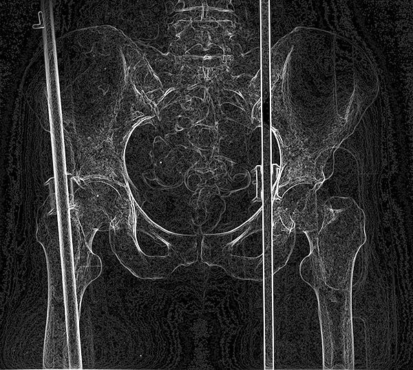
These cases can be reviewed individually or with other students before or after mini-consolidation session on Pelvic Imaging.
For a brief review of types of radiological imaging, with narrated slides, click this LINK.
Learning Objectives:
After review of online resources and in class mini-consolidation, students will be able to:
1. List most appropriate imaging modalities for the uterus and prostate, and recognize images from each modality including CT, MR and US.
2. Explain why a congenital abnormality on an image of the kidney could relate to findings in the uterus using your understanding of embryology, and list one interventional procedure that can be used to evaluate uterine configuration and one for evaluation of patency of the vas deferens.
Imaging Anatomy Pelvis Case 1 Imaging Anatomy Pelvis Case 2