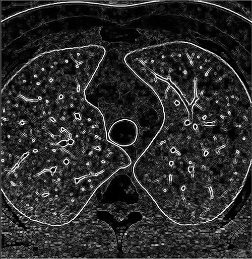
After completion of this self-directed learning module on Imaging of Lungs and Pleura, students will be able to:
- Recognize and describe the following findings on frontal chest radiographs: normal vs increased or decreased lung volume, normal vascular distribution in the upright position, normal bronchi, and normal pleural surfaces including costophrenic angles
- List three areas related to lungs and pleura that are better seen on the lateral chest radiograph than the frontal.
- Describe the parts of the secondary pulmonary lobule that can be seen on chest radiography and CT in normal vs abnormal situations, such as Kerley B lines or tumor infiltration of interstitium.
- Recognize and decribe, using appropriate imaging terminology, features of abnormal lung on CT, including morphology of interstitium, bronchi and vessels, and indicate what technical parameters are important in visualizing each of these areas on CT.
